


概要
滲出性AMDは治療可能な疾患であり、効果的な治療法の開発が現在も続けられています。以前は、レーザー光凝固術という治療法でしか滲出性AMDの漏れを治療する事はできませんでした。90年代に入ってからは、ビスダイン(血流に直接注射し、目の患部に照射したレーザーによって活性化する薬)を使用した光線力学治療(PDT)が開発されました。
2004年以降、標的治療とよばれるより効果的な治療法が開発されました。これらの治療法は、異常な毛細血管の成長を促す特殊なタンパク質であるVEGFの働きを阻害します。抗VEGF療法は滲出性AMDに対する画期的な治療法であり、世界中の多くの患者をこの病気から救っています。

臨床成果
血管新生のプロセスをターゲットにした治療は総称して抗血管新生療法と呼ばれます。また、眼球内の血管新生を引き起こすタンパク質であるVEGFの働きを阻害するため、抗VEGF療法とも呼ばれます。抗VEGF療法はAMDの進行を遅らせ、視力の回復に至る場合もあります。しかし、この治療法は瘢痕組織が形成され、不可逆的な視力低下がはじまる前にのみ効果を発揮します。
抗血管新生療法
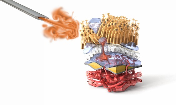
VEGF(血管内皮細胞成長因子)は、正常な血管の内側を覆う内皮細胞を活性化させ、血管新生を促進するタンパク質です。
滲出性AMDではVEGFが網膜内で過剰に発生し、黄斑下部の異常な血管新生を促します。これらの血管は非常に脆く、眼球組織に漏出した体液や血液は黄斑を損傷させ、中心視野を失う原因となります。
抗VEGF療法はVEGFタンパク質を標的とし、その発生を抑制します。この治療により新たな血管の成長を阻害し、体液の漏れや出血を事前に防ぎ、健康な視力を守ります。
現在、滲出性AMDの治療に有効と証明された抑制物質は4種類存在します。
- マキュゲン(ペグ化抗VEGFアプマター)― 滲出性AMDの治療薬として最初に認可を受けた抑制物質。
- ルセンティス(ヒト化抗VEGF抗体)- 滲出性AMDに良く用いられている治療薬。
- アイリア(VEGFトラップ)- 最近米国で認可された治療薬で、2か月に1回の頻度で済む。日本でも2012年に認可されたため、保険が適用されます。
- アヴァスティン(ヒト化抗VEGF抗体)- 抗VEGF作用のある抗ガン剤で、滲出性AMDの治療として網膜専門医による適応外使用が行われている。
ここに挙げた抗VEGF剤はすべて、網膜専門医により直接目に注射されます。網膜専門医はこのような治療を痛みやリスクを最小限に抑えて行う訓練を受けています。治療の具体的な頻度は患者の病状に基づいて網膜専門医が決定します。
網膜専門医により行われた場合、抗VEGF療法は比較的安全な治療法であるとされています。しかし全く副作用が現れない訳ではなく、リスクと治療の効果のバランスを取りながら使用する事が大切です。抗VEGF療法には以下のようなリスクがあります:
- 眼感染症
- 眼圧の上昇
- 網膜剥離
滲出性AMDは、生涯に渡る経過観察と治療が必要な慢性疾患です。現在使用可能な抗VEGF療法を用いた場合、視力の保全や血管新生の抑制の為には定期的な治療が必要となります。網膜専門医の指示通りのスケジュールで治療を行わなかった場合には、視力低下や永続的な失明につながる恐れがあります。治療の頻度や期間は担当の網膜専門医と相談して決定してください。
参考文献
マキュゲン
マキュゲンは滲出性AMDの治療のために開発された、VEGFを標的とするアプマター(短いヌクレオチド鎖) で、眼球に直接注射されます。マキュゲンは視力低下を抑えますが、視力回復効果はありません。マキュゲンは6週間に1回、0.3mgずつ投与されます。
滲出性AMD患者1200人を対象とした研究によると、マキュゲンを投与された患者の半数以上は、研究開始から1年間における視力低下が視力表で3段階以内に抑えられる事が分かっています。また、マキュゲンを投与された患者の約65%で視力の安定が認められています。
報告事例の多い副作用は、目の炎症、視界のぼやけ、眼球結膜出血、目のかゆみ、目の痛み、浮遊物、眼圧の上昇です。眼球への注射に伴う深刻な副作用としては、眼内炎や網膜剥離が挙げられます。
参考文献
ルセンティス
ルセンティスは網膜疾患の治療を目的として開発された、モノクローン抗体フラグメントと呼ばれる抗VEGF剤です。眼球に直接注射され、視力を安定または改善する作用があります。ルセンティスのパッケージには月1度、0.5mgの投与が最も効果を期待できるとあります。網膜専門医の中には、1か月に1回よりも少ない頻度でルセンティスを投与する医師もいます。
1300人以上を対象とした臨床試験の結果、0.5mgずつの投与を月1回のペースで2年間行った場合、患者の約90%において視力を安定(顕著な悪化が見られなかった)させる効果がありました。また、患者の約30%において著しい視力回復が見られました。
報告事例の多い副作用は、眼球結膜出血、目の痛み、浮遊物、眼圧の上昇、そして目の炎症です。眼球への注射に伴う深刻な副作用としては、眼内炎や網膜剥離が挙げられます。
参考文献
アイリア
アイリアは抗VEGF剤の中でも融合タンパク質と呼ばれるもので、眼球へ直接注射します。アイリアは、VEGFだけではなく胎盤成長因子(PIGF)と呼ばれるタンパク質にも作用しますが、PIGF も滲出性AMDの患者の網膜に過剰に存在することが分かっています。1か月に1回の投与を3回行ったのち、2か月に1回の投与に頻度を落としてもルセンティスを月1回投与した場合と同様の効果を得ることができます。
滲出性AMD患者約2400人に対して行った臨床試験において、ルセンティスを月1回0.5mgずつ投与される患者と、アイリアを最初の3か月間のみ月1回2mgずつの投与されたのち、2か月に1回同量を投与される患者との比較が行われました。1年間の治療の結果、2か月に1回投与されるアイリアは1か月に1回投与されるルセンティスと同様の視力安定もしくは回復効果を発揮する事が分かりました。また、安全性においても同程度の結果が得られました。アイリアはより少ない回数の注射で効果を得ることができ、体への負担も少ないといえます。
報告事例の多い副作用は眼球結膜出血、網膜出血、そして視力の低下です。眼球への注射に伴う深刻な副作用としては、眼内炎や網膜剥離が挙げられます。
参考文献
Beovu
Beovu is a small-sized, single strand antibody fragment with enhanced tissue penetration and active drug binding, that delivers a high concentration of the anti-VEGF drug to the target area in the eyes of patients with wet AMD. Beovu allows for 3-month dosing intervals after a 3-month loading phase, which may offer benefit to patients on other anti-VEGF drugs that require four week dosing periods, and for which frequency of monthly injections creates a burden. In two Phase III clinical trials, Beovu was effective in half of the patients at a dosing interval of 12 weeks in between treatments; the other half required treatments at 8-week intervals.
On average, patients gained seven letters on the eye chart, and 30 percent of patients gained at least 15 letters. In the trials, Beovu given every 2-3 months was non-inferior to Eylea given every two months. In addition, those patients receiving Beovu had reduced central retina thickness, and less retinal fluid detectable by OCT imaging.
References
適応外の治療
アヴァスティン
アヴァスティンはもともと抗ガン剤として開発された薬ですが、抗VEGF作用があります。日本では、滲出性AMDに対するアヴァスティンの使用は認可されておらず、保険は適用されません。
Implantable Telescope
(English) Implantable Telescope
The Implantable Miniature Telescope (IMT) was FDA-approved in July 2010 for end-stage AMD. This device is manufactured by VisionCare Ophthalmic Technologies Inc. of Saratoga, California, and its approval followed a 219-patient, multi-center clinical study in which 90 percent of patients achieved at least a 2-line gain in either their distance or best-corrected visual acuity, and 75 percent their level of vision from severe or profound impairment to moderate impairment.
Patients 75 years or older with stable to severe profound vision impairment due to blind spots (bilateral central scotoma) are now eligible for surgical implantation of this device, which projects images at greater than two times magnification onto the retina to improve central vision. Options are available for 2.2 and 2.7x magnification. Pre-training with an external telescope with a low vision specialist is required prior to procedure to ensure the device can have a benefit, as well as to determine eligibility (inadequate peripheral vision). Post-op visual training is required, as well.
One risk of this device includes the loss of corneal endothelial cells, which are essential for maintaining corneal clarity. The degree of this loss can be chronic (up to 5 percent per year). Major losses may have negative downstream effects, including corneal edema (swelling), decompensation (further loss of function), and ultimately a need for corneal transplant. 10 eyes in the above named study had corneal edema (unresolved), half of which resulted in corneal transplant. Calculated 5-year risks for adverse outcomes were calculated as follows: corneal edema (9.2 percent), corneal decompensation (6.8 percent) and corneal transplant (4.1).
Two post-approval studies are being carried out to further delineate risk, including a two-year follow up of the study’s initial two year cohort as well as a novel study of 770 newly enrolled subjects (focusing on endothelial cell density and related sequelae). Measures were also taken to ensure patients are informed of risks, including detailed labeling (manufacturer and FDA-generated) and an Acceptance of Risk and Informed Decision Agreement.
Visudyne
(English) Visudyne (photodynamic therapy)
Photodynamic therapy with Visudyne injection was FDA-approved in 2000 for the treatment of age-related macular degeneration (predominantly classic subfoveal choroidal neovascularization). Alternate indications include pathologic myopia or presumed ocular histoplasmosis. This drug is activated by light, and functions to block the mature vessels that may be expressing less or no VEG-F, contributing to persistent AMD activity in spite of anti VEG-F treatments.
The most common adverse events reported with Visudyne include injection site reactions, blurred vision, decreased visual acuity and visual field defects (10-30%). Contraindications include porphyria or known hypersensitivity to any component of the drug formulation.
Macular Translocation Therapy
(English) Macular Translocation Therapy
Macular translocation therapy is a surgical intervention for wet AMD only, and involves detaching and rotating the retina, such that placement will allow the macula to sit on a new, healthy base. It is not used for dry AMD due to degeneration occurring in the retina’s new position. Eligibility entails central vision loss in both eyes, with one having developed the condition in the preceding 6 months. It has been shown to be effective for some when performed promptly.3
Complications include cataract formation, infection, intraocular bleeding, retinal detachment and/or tears and total vision loss. Since retinal rotation causes tilted or double vision, a second correction surgery for eye muscles is required 2 months after the initial procedure. Both are performed on an outpatient basis.
References